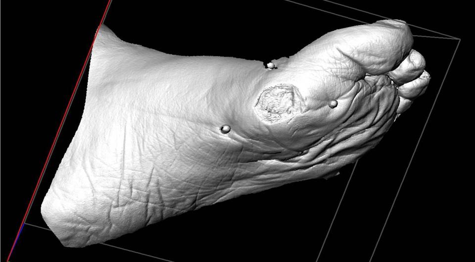
3D camera for monitoring foot ulcers
Diabetes is a disease that can cause deep foot ulcers that take a lot of time and resources to heal. Diabetic foot ulcers are among the most serious and cost-consuming complications in relation to diabetes and often lead to amputations.
PROJECT PERIOD
Start: 2016
End: August 2019
Numerous studies have shown that the size of the ulcers, including the depth, is one of the essential factors in delayed healing. It is important to monitor the wound healing process in order to change the treatment plan in the early process.
The Region of Southern Denmark has tested a telemedicine service for treatment and monitoring of foot ulcers, where a nurse treats the patients at home, so the patients can avoid many trips to the hospital. The nurse could take pictures of the ulcer and share them in an online platform where wound specialists can review them and provide guidance on the treatment of the ulcer. To improve the telemedicine service, a 3D camera has been developed alongside the test of the telemedicine service to provide better monitoring of the ulcers, as the 3D camera can measure the depth of the wound, not only the surface of the wound that a 2D image can capture.
AIM
The PhD project set out to perform a clinical validation of the existing prototype of the “3D-Wound Assessment Monitor (WAM)” for monitoring of diabetic foot ulcers. The project compared the 3D measurements with standard measuring methods (2D method and gel injection).
The aim of the project was to clarify if the 3D pictures of ulcers could provide more accurate measures to improve the wound care and increase the healing rate of the ulcers. The future vision is that the 3D camera can be useable for both the telemedicine service in the primary sector and also at the hospital’s specialised center for wound treatment.
RESULTS
The project showed that the camera is an accurate and reliable method for measuring wounds in three dimensions, particularly in wounds with an area above five cm2.
The study monitored the wound healing of 150 diabetic foot ulcers using the 3D-WAM camera and compared them to the traditional 2D image method during a period of eight weeks. It turned out that the wounds healed significantly faster estimated from the changes in 3D area measurements compared to 2D area measurements. This can be explained by the fact that the changes in 2D area (surface area) is only an expression of the wound healing from the surface of the wound, whereas the changes in 3D area is an expression of both the wound healing from the wound bed and the surface of the wound. However, in small wounds with an area below five cm2, the study found that the difference between the changes in 2D area and 3D area measurements were very small because the wounds are often shallow.
With a 3D camera has an accurate and objective tool, a specialist in the hospital will be able to provide better guidance for the in-home treatment of diabetic foot ulcers.
The 3D camera (Wound Assessment Monitor (WAM)) used in the study was a prototype developed by the company Teccluster and is not yet available on the market. For this reason, the WAM technology will not be implemented as part of the diabetes care at OUH at this point in time. However, this PhD study has proven that there is a clinical potential to further develop the technology and make it market-ready in terms of both design, functionality and price.
PARTNERS
The project was a PhD project by Line Bisgaard Jørgensen. View Line’s publications.
The project was a continuation of a PhD project by Benjamin Rasmussen where the idea for the 3D camera and an early prototype was developed.
Line Bisgaard Jørgensen
MD, PhD
Odense University Hospital, Department of Internal Medicine and Acute Medicine
(+45) 6591 9653 line.bisgaard.joergensen@rsyd.dk

Knud Bonnet Yderstræde
Senior researcher, MD, associate professor
Department of Endocrinology, OUH & Department of Clinical Research, University of Southern Denmark
(+45) 6541 3427 knud.yderstraede@rsyd.dk

